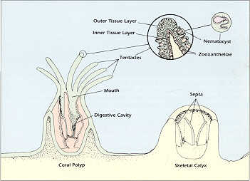
Figure 1. Diagram of coral polyp and calyx structures
.
What is a Coral?
What we commonly call corals includes a variety of types of organisms that biologists refer to as coelenterates or cnidarians. One of the main physical characteristics of this group is that they all have a single body cavity and opening, a coelenteron, that doubles both for the ingestion of food and for the release of digested wastes. Another characteristic is that corals and other coelenterates have stinging cells, or nematocysts, that are normally carried within special cells on the animal's surface-. When potential food prey is present in the water, the coral ejects these stinging cells to entangle or poison the prey, which the coral can then consume.
Figure 1. Diagram of coral polyp and calyx structures
.
The body structure of corals and their close relatives, the sea anemones, appears as an upward facing, radially or biradially symmetrical polyp. In each polyp the animal's mouth lies in the center of a ring of tentacles that surround the perimeter of the oral disk. The nematocysts are most abundant on the surface of these tentacles, which can elongate dramaticaJly when the coral is actively feeding. Within the body cavity, digestion is accomplished on the surfaces of specialised filaments or mesenteries, which secrete enzymes that quickly reduce ingested prey to its components. Most corals are, therefore, potentially efficient predators, although many types seem to have developed other means of meeting their energy requirements (see below).
Although the term "coral" is often used in a general sense, it usually refers to reef or hard corals, which are more formally called scleractinians. These are the corals that may form large coral heads and which are the primary building blocksof coral reefs in tropical oceans: The common characteristic of. this group is the secretion of a basal skeleton of calcium carbonate as the mineral aragonite. The livng portion of thIs group usually has its radial symmetry divided into multtiples of six, which is most apparent in the numbers of tentacles that surround each polyp. This pattern is also shown in the pattern of the calyx, (plural=calyces) or cup, which is secreted by each polyp, and which remains after the live tissue is cleaned from a coral skeleton. Each calyx is divided by a number of septa, again usually in multiples of six, which look like sharp ridges radiating from the center of the calyx.
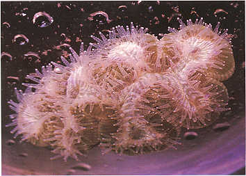
A reef coral can be a single polyp and calyx, but in most cases reef corals grow as colonies with hundreds, even thousands of polyps and calyces on the same coral skeleton. The variety of forms that are created by this colonial growth make up some of the most beautiful structures to be found in nature. These structures can range from delicate, branching bushes and arbors, to robust tables, intricate flower-like leaves, and multiple starbursts to branching fingers and massive boulders that have brain-Iike fissures on their surface. The variety of shapes is determined by the pattern of budding of new polyps from older polyps as the coral grows, and whether the polyps become separated or continue to share a common mouth within the original ring of tentacles. The calcium carbonate skeleton also varies substantially in density and strength according to the coral species. Some corals have very dense skeletons and can resist a great deal of wave disturbance, while others are light and porous and are restricted to the calm waters of embayments. However, these porous corals have the advantage of having faster growth rates and may have live tissue penetrating the coral skeleton. This tissue within the skeleton gives these corals a greater ability to recover from stresses which may kill off the coral surface tissue.
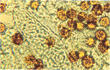
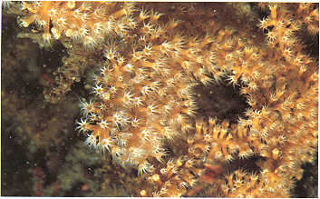
Because of their flower-like appearance, corals were classically referred to as "zoophytes", indicating that they were recognized as animals, but had many external characteristics of plants in their patterns of growth. Now we know that this original descriptive term was quite valid, but for a different reason. Coral biologists have long recognized that all hard corals with rapid growth rates contain massive numbers of single celled algae called zooxanthellae within the cells of the coral tissue. These algal cells appear under the microscope as yellowish-brown spheres, and they are extremely abundant in the coral tissue, normally numbering millions of cells per square centimeter of coral surface. The zooxanthellae provide the major coloration of most living hard corals and many soft corals, which usually appears as shades of brown or green.
The actual functional relationship of these internal algae to the coral has been studied intensively and was a subject of controversy for many years. Coral biologists always recognized that the presence of the intracellular algae was necessary for hard corals to achieve the rapid growth rates which enable them to reach massive sizes and form coral reefs. Most non-reef forming or solitary corals, although they have similar calcium carbonate skeletons, do not contain zooxanthellae and do not grow to large sizes. The question remained as to how the association between the corals and their zooxanthellae might aid or benefit either of the partners? When rapid growth occurs, an animal produces metabolic carbon, phosphorus and nitrogen wastes that must be removed or they will poison the growth process. Corals, being very simple organisms, have no specialized structures for such waste removal. However, the abundant zooxanthellae within the coral tissue provide a mechanism for removing wastes and therefore permit rapid coral growth. The waste products produced by the coral tissue are the raw materials used for photosynthesis by the zooxanthellae and thus are kept from rising to toxic concentrations. The zooxanthellae, on the other hand, benefit by having a ready supply of nutrients for photosynthesis. This is an example of what is termed mutualistic symbiosis, where both partners benefit from being joined in a close association.
For many years, this removal
of waste products was considered to be the only benefit to the coral provided
by the algae. However, the question remained whether the organic matter
produced by the zooxanthellae could be utilized in any way by the coral
tissue, i.e. was the plant partner of the association producing food for
the animal? Research in the past twenty five years has verified that
such a nutritional relationship does exist. A major portion of the energy
fixed by the zooxanthellae passes directly to the animal tissue in a highly
usable dissolved form. Coral therefore do "feed" on their zooxanthellae,
although not by conventional means.
The term "corals" often includes other varieties of coelenterates that have somewhat different characteristics than scleractinian corals. The largest of these groups are the octocorals which includes both hard and soft forms and the gorgonians or horny corals. Their common characteristic is that, instead of being in multiples of six like the hard corals, the structures of their polyps, such as tentacles, grow in multiples of eight. The tentacles of these octocoral polyps are pinnate or feather-like. The most common hard octocoral is the organ pipe coral, which grows from a deep red skeleton which is formed by parallel rows of tubes. In life, however, this coral shows the white coloration of its polyps and feathery tentacles, which resemble thousands of small white flowers.
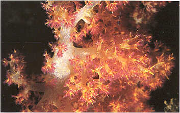
The soft octocorals have many features in common with the hard corals. Most grow as upwardly facing polyps, contain symbiotic zooxanthellae and have tentacles and nematocysts that may be used in food capture. The primary difference is that the soft corals do not lay down an external hard skeleton that remains as a permanent structure after the coral dies. Instead, the soft corals get their structure and body support from calcium carbonate spicules that are deposited within their body walls. These corals may look like quite drab leather-like gray-green sheets on the reef surface, or they can be very colorful and beautiful, such as the dendronepthid or teddy bear corals which have a crown of red to orange tentacles on a translucent white stalk.
The gorgonian octocorals grow in a
variety of forms such as elaborate sea fans and sea whips. Their supporting
structures, instead of being external calcium carbonate skeletons or internal
spicrules, are rods of horny material that form the backbone of each net-like
fan or branching whip. The living tissue and polyps grow on the surface
of this rod. Sea whips are generally quite small, but sea fans can grow
to two or three meters in diameter, forming a large net of feeding polyps
that make up an efficient network of plankton feeders. These forms
depend on zooplankton for food, rather than organic matter fived by internal
zooxanthellae. Therefore, they are usually most abundant where they
have clean hard surfaces to attach themselves to, and in areas of moderate
to strong currents where plankton are abundant.
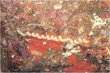
Plate 6. Specimens of a gorgonian purple sea fan on
the reef showing partly expanded polyps.
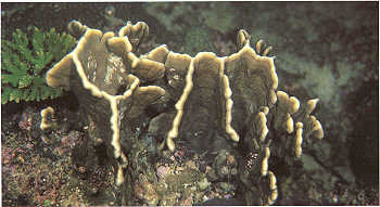
Fire coral is abundant in most areas in the tropics, but in Oman it is only found in the Dhofar region. Fire coral belongs in yet another coelenterate group called the hydrozoa. many of the toxic and stinginging animals such as the Portuguese Man O' War found in ocean waters are also members of this group. Fire coral's calcium carbonate, basal skeleton grows in a wide variety of shapes, and it is always a rich yellowish brown in color. Fire coral is distinct from all other corals in that its calyces appear as pin holes with neither septa nor tentacles being visible. The appearance of these pin holes is the source of fire coral's scientific name, Millepora. The potency of its capability as a predator is confirmed by the painful stings that it will inflict on a person who comes in contact with its surface and nematocysts.
The final group of animals that are generally referred to as corals are the precious corals. The term "coral colored", classically refers to the color of deep living; pink precious coral which has been used as jewelry and treasure since ancient times. Of somewhat similar appearance is a certain type of hydrozoan called Stylaster, which is actually more closely related to fire coral and occurs in shallow water, but is of no commercial value. Pink precious coral ranges in color from deep red to pale pink. It is the hard internal skeleton of a type of gorgonian that usually occurs deep in the ocean to thousands of meters, but may be found in the Mediterranean Sea as shallow as 100 meters. A closely related, equally beautiful type is gold precious coral, which also is a deep living gorgonian. Traditionally taken as a by-product of deep water fishing with tangle nets, these precious corals have been harvested in more recent years using deep water submersibles.
Black coral is also used commercially in making jewelry and small sculptures. Black coral trees generally occur in intermediate depths within the range of SCUBA divers who may harvest the trees for profit. The trees can grow up to several meters in height in areas where currents are strong and plankton food plentiful. While alive, black coral may appear white, yellow or even red depending on the color
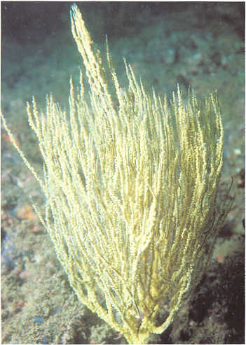
of the live polyps which cover the internal skeleton, similar to gorgonians. Cleaning off the live tissue reveals a hard black, somewhat spiny skeleton which, when ground and polished, shows a beautiful shiny surface, as do pink and gold corals. Another close relative of black coral, wire coral, has a similar structure of polyps growing on a supporting rod, which in this case is tightly coiled and can be several meters long. However, wire coral never grows sufficiently thick to be used in jewelry making.
The scleractinian hard corals are
the most common and abundant coral type to be found on most coral reefs
in the tropical world and in the coral growing areas of Oman. Certain types
such as gorgonians and black corals are relatively uncommon in Oman. and
others such as organ pipe coral and fire coral occur only in the Dhofar
region. This book will therefore concentrate on the hard and soft corals
which are abundant in Oman's nearshore waters and which are easily available
to be seen by snorkelers and SCUBA divers. Before these corals are described.
the coral communities of Oman will be reviewed in the context of their
relationship with reef corals that occur in the Indian and Pacific Oceans.